ORIGIN OF SWEET SPOT™
The SWEET SPOT™ was created for two main reasons. The first was to improve patient safety by decreasing the complications caused by improperly positioned venous access devices (VAD) as encountered by me and other members of my IR practice. The second was to remove the always subjective and sometimes erroneous chest x-ray (CXR) interpretation of VAD tip position (as well as resultant unnecessary recommendations for repositioning) in my own large and highly sub-specialized radiology group (103 members as of 2013).
DEFINITION OF SWEET SPOT™
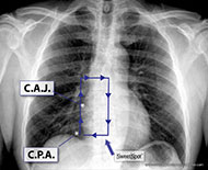
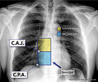
The SWEET SPOT™ is a rectangular template superimposed on a frontal CXR, whose margins and internal area are acceptable for VAD catheter tip position. This is tailored to the individual patient's chest x-ray. As such, it has no fixed length or width but does have fixed proportions with the craniocaudal length being twice the width on a frontal chest x-ray. Depending on patient anatomy, it can exceed 8 x 4 cm. The template (see Figure 01) is simply compared in size to the CXR at hand. This template is very easy to memorize and just as easy to teach.
The SWEET SPOT™ also has a fixed center point. It is centered over what I believe is the most accurate radiographic estimation of the cavo-atrial junction, i.e. on a frontal CXR, the initial outward bulge of the lower right cardiomediastinal margin due to the transition from the smaller, more tubular superior vena cava to the more capacious and rotund right atrium. It was designed to be twice as wide as the lower third of the SVC to allow for the curving course of many catheter tips as they enter the right atrium - especially with left-sided access. The SWEET SPOT™ extends from the lower third of the SVC to the most inferior extent of the right atrium - stopping at the frontal CXR's cardiophrenic angle (see Figure 01).
The SWEET SPOT™ is the best anatomically based and radiographically confirmed practical approximation of the cavo-atrial junction. It is independent of patient factors such as age, size, patient position for chest x-ray, lung volume or type of VAD. It also encompasses the various leading societal recommendations for catheter tip location (see below). As such, it allows for the necessary significant flexibility in acceptable VAD tip location based not only on patient, chest x-ray and catheter variables as just mentioned but also inserter variables such as societal affiliations, personal experience and personal convictions.
CLINICAL USE OF SWEET SPOT™
It was initially adopted practice wide by my group — Inland Imaging, Spokane, Washington in 2007 as the sole acceptable chest x-ray standard for VAD tip location - which it still is. As such, it also became the standard of acceptability for our recently completed Sherlock 3CG trial.
In this same trial, the vast majority of the Sherlock 3CG guided catheter tips — whose technology targets the S.A. node, were at the center of the SWEET SPOT™, i.e. the radiographic cavoatrial junction, thus confirming both chest x-ray and ECG methods of catheter tip verification. The two methods, therefore, cross-validate and have the same end point — optimal VAD tip location.
SOCIETAL RECOMMENDATIONS FOR V.A.D. CATHETER TIP LOCATION
In 1989, the US FDA (Task Force FDA Drug Bulletin 1989:15) issued an advisory statement that it is unacceptable for catheter tips to be placed or permitted to migrate into the heart for fear of cardiac tamponade.1 During this time, other studies provided evidence that catheter tips placed high in the SVC or outside the SVC increase the risk of thrombus and catheter malfunction. (Kearns2, Caers3, Cadman4). In 1998, the National Association of Vascular Access Networks (NAVAN), now "AVA," published a position statement recommending that the “most appropriate location for the tip of PICCs is the lower one-third of the SVC, close to the junction of the SVC and the right atrium” and not extending into the right atrium. (NAVAN tip position).5 In 2006, the Infusion Nurses Society updated their Standards of Practice and again in 2011 stating that “CVC tips should be located in the lower third of the SVC to the CAJ” (INS 2011).6 While the authors and organizations cited above favor positioning outside the right atrium, the Society of Interventional Radiology (SIR) states (in their Quality Improvement Guidelines for Central Venous Access) that the tip should be “in the cavoatrial region or right atrium” (2010 SIR Dariushnia)7 and this view is also stated in the European Society of Parental and Enteral Nutrition (ESPEN) Guidelines (Pittiruti 2009).8 Historically this inferior migration of catheter tip position acceptability is coincident and not unrelated to manufacturers developing V.A.D.'s made out of softer materials.


